Students will learn about the importance and principles of radiological imaging and diagnostic technology used in diagnostic of diseases and injuries of teeth and maxillofacial area. They will get acquainted with the digital X-ray, computer tomography, intervention radiology, diagnostic ultrasound and magnetic resonance. They will get knowledge of the indications for each recording technique and knowledge of the intraoral imaging technique. They will adopt general principles for the analysis and interpretation of X-rays and other radiological studies.
Normal radiological anatomy of teeth and periodontal tissues, upper jaw, lower jaw, jaw hinge and other maxillofacial areas. Pathological-anatomical changes, important for understanding radiologically visible signs. Radiological characteristics of congenital abnormalities, injuries, infections, tumors and degenerative changes in teeth and periodontal tissues, upper jaw, lower jaw, jaw hinge, and other maxillofacial areas. Clinical indications for the use of certain radiological technology, intraoral imaging technique and image development.
Required course materials:
1. Dentalna radiologija. S. Janković, D. Miletić. Medicinski fakultet Split
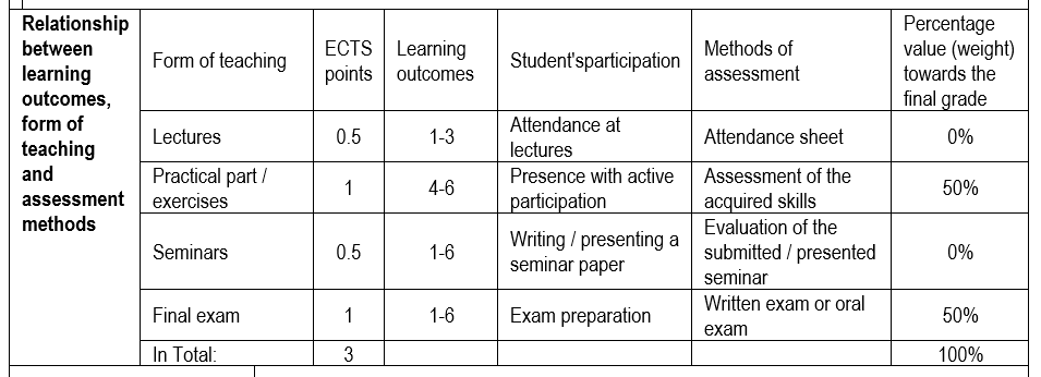
Upon completion of this course, students will be able to:
1. To determine radiological terminology and acquire the ability of radiological analysis of anatomical structures of the upper and lower jaw and pathological processes of teeth.
2. Integrate the radiological picture with pathoanatomical changes.
3. To determine the characteristic radiological manifestations of diseased conditions of the jaw.
4. Select an imaging diagnostic modality suitable for the diagnosis of pathological changes.
5. Argue the choice of radiological diagnostics and radiologically integrate radiological terminology with the clinical finding.
6. Critically evaluate the choice of radiological modality with the aim of preventing excessive exposure to ionizing radiation or undesirable and harmful effects.


